November 2019 Update
Louise Ludlow has kindly provided CIKA with the following report on the operation of the tissue bank at the RCH.
Working together – The Children’s Cancer Centre Tissue Bank and Anatomical Pathology
The timely procurement and processing of tissue samples after surgery is crucial. After the tissue is surgically resected, the changes in environmental conditions such as temperature and oxygen levels can lead to rapid degradation rendering the sample useless. Therefore maintaining tissue integrity by avoiding delays in obtaining the specimen is of high priority
Following removal in theatre, the specimen is transported directly to the Anatomical Pathology service at The Royal Children’s Hospital for testing. Scientists and pathologists work quickly and efficiently to provide a diagnosis which is relayed to the surgical team. If excess tissue exists, the scientists in Anatomical Pathology page the team at the Children’s Cancer Centre Tissue Bank, which is housed at the Murdoch Children’s Research Institute. Staff from the Tissue Bank then collect and process this tissue for banking. This may occur after hours, or on the weekend where collection and processing of tissue is facilitated by our rostered scientists.
The Tissue Bank then relies on Anatomical Pathology to validate the tissue that we bank. This involves the scientists preparing a frozen section for the pathologist to assess by microscopy and confirm that our banked tissue matches the diagnostic report. This is an important check to ensure we supply the correct tissue for our research projects
Anatomical Pathology plays an integral role in providing samples to the Children’s Cancer Centre Tissue Bank. Duncan MacGregor (Director, Anatomical Pathology), Bronwyn Christiansen (Principal Scientist) and their team have been assisting our Tissue Bank with this process for the past six years. This key relationship and their in-kind support is essential to the success of our Tissue Bank.
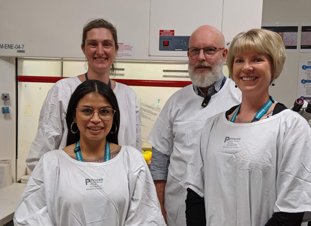
From left to right back row: Bronwyn Christiansen (Principal Scientist) and Duncan MacGregor (Director, Anatomical Pathology).
From left to right front row: Elena Fernandez (Tissue Bank Research Assistant) and Louise Ludlow (Tissue Bank Coordinator).
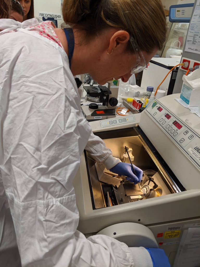
The section is then mounted onto a microscopic slide for staining and assessment.
You must be logged in to post a comment.