Research Report 2016
Dr Louise Ludlow was the guest speaker at The April Macedon Ranges Focus Group luncheon on 8 April 2016. Louise established and now manages the Children’s Cancer Centre Tissue Bank and her talk centred around her background and the research she has undertaken at the Murdoch Children’s Research Institute. Here is a transcript.
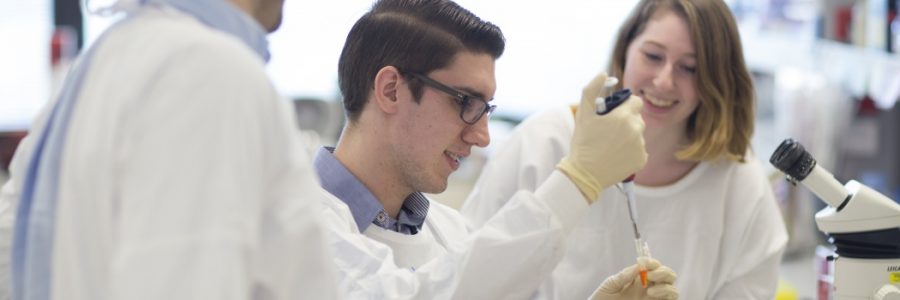
I started working at the Murdoch Children’s Research Institute three years ago in the role of Tissue Bank Coordinator. I come from a research background having completed my PhD at The Peter MacCallum Cancer Centre in 2005. I studied a family of genes involved in cancer immunology. It was five years of hard work and I enjoyed the excitement of making new discoveries. After completing my PhD I took up a three year role at Northwestern University in Evanston just outside Chicago. Here I investigated the ability of viral proteins to escape the immune system. The research environment was competitive and work ethic was fierce. It was a wonderful life experience but terribly cold in the winter!
Upon return to Australia I took up a four year position at The Burnet Institute and The University of Melbourne investigating the immune response to malaria and HIV infection. In my experiments I used blood cells collected in PNG and discovered the incredible value of using banked tissue.
My
role as Tissue Bank Coordinator has
provided a fantastic opportunity to expand upon my laboratory skills
and to meet dedicated and inspirational people like yourselves. The
role has provided many challenges such as gaining our ethical
approval to start the Tissue Bank, communicating with gruff surgeons
and overworked oncologists!
Our Tissue Bank has been in
operation for two years. During this time we have consented just
under five hundred patients and banked over a thousand samples. We
have banked many rare and interesting solid tumour specimens along
with blood and bone marrow samples.
Tissue banking is a highly
collaborative process requiring great levels of communication and
networking. Over fifty staff including consultants, surgeons,
haematologists, oncologists, specialist pathologists, researchers and
a roster of on-call scientists ready to receive and process tumour
material on any day at any time make up this team. Our process begins
with consenting the patients and their families in the clinic or
bedside. We then collect and process the samples in our laboratory on
Level 5 South.
All research projects investigating childhood
cancer rely on using tissue samples removed from patients in the
operating theatre or in the clinic during the normal course of
clinical investigation and treatment.
The role has provided
experiences that have put life in perspective. Meeting the mother and
brothers of a young girl who passed from a rare and incurable brain
tumour. Her capacity to donate this tumour for research. My joy is
showing this family the immortalized cell line under the microscope
which I generated. Knowing this tumour tissue and cell line will be
used to make a difference in the fight against these brain tumours.
Seeing the amputated hand of a child containing a large muscle
tumour. Knowing that this tissue we bank will make a
difference.
Through the devastation and unbearable grief this
disease causes there is a shining light that is research. Our Tissue
Bank is an incredibly important resource for cancer research and is a
core part of the Children’s Cancer Centre. Our vision is that
through research we can not only improve treatment but contribute to
implementing a personalised model of care within the RCH. Banked
samples are already being utilised in a number of research studies
being conducted on campus and samples have also been dispatched to
contribute to collaborative international studies.
I
would now like to take the opportunity to explain two projects that
have used samples provided by the Tissue Bank.
The first
study involves
investigating pilocytic astrocytoma and is the work of a PhD student
Alex Sexton-Oates working with Prof., Richard Saffery. PA is the most
common brain tumour in children under the age of eighteen. In fact
one in five children diagnosed with a brain tumour have a PA. PA has
an excellent survival rate and treatment consists of surgical removal
of the tumour. Radiotherapy and chemotherapy may be given in cases
where the tumour cannot be completely removed. Extra treatment may
also be given if the PA grows back after surgery, this is called
‘recurrence’ and happens is up to 30% of children.
Children
may be left with long-term negative effects on their brain function
and mental health. These long-term effects are due to both the
treatment children receive and the location of their brain tumour.
The research is asking two questions. What is different about PAs
that grow in difference parts of the brain? What is the difference
between PAs which do not recur after surgery and those which
do?
Alex used a new technique called a methylation array to
analyse a group of PA. Through this technique she was able to
identify the tumours which recurred. This work will lead to
identifying new treatments for the more aggressive PAs and the
ability of predict at diagnosis which children will need to return
for regular brain scans.
The
next project involves
understanding chemotherapy-induced heart disease. We have processed
nearly three hundred blood samples toward this project which is being
led by Dr Rachel Conyers, an oncologist together with Dr David
Elliot, an MCRI researcher.
A side effect of a number of
valuable chemotherapeutic drugs is heart damage. Cancer survivors
treated with these drugs are nine times more likely than average to
develop heart failure. However, only 20-30% of patients are
susceptible to chemotherapy-induced heart failure. The aim of this
research is to find out why some patients are resistant and some are
sensitive to this toxicity. The study aims to genetically profile
over 150 childhood cancer survivors and develop stem cell technology.
The goal is to set up a clinically applicable tool to predict patient
sensitivity to chemotherapy. This will enable a more tailored
chemotherapy to patients resulting in less cardiac toxicity.
The
stem cell technology involves taking the white cells from peripheral
blood and reprogramming them to an embryonic stem cell-like state.
These cells are then differentiated to cardiomyocytes which beat in
culture.
There are two additional aspects of the Tissue Bank:
We
generate neurosphere cell lines from high grade brain tumours. These
cell lines represent valuable tools for understanding tumour biology
and for testing novel drugs before their use in children.
The
CCC has an active research arm in international collaborative large
scale clinical trials. The CCC Tissue Bank provides tissue processing
to enable patient participation in international clinical
trials.
MCRI is a great place to work! The director
of the CCC, Francoise Mechinaud, has been the driving force behind
establishing the bank and Prof., Richard Saffery provides guidance
and advice. There are many interesting areas of research, many
dynamic and dedicated researchers. Some areas of research include
expansion of genomics for early diagnosis and improved treatment,
rotavirus vaccine work, development of an inhaled vaccine
device.
Much of this research at MCRI could not be carried out
without philanthropic funds. We are extremely grateful for your
support.